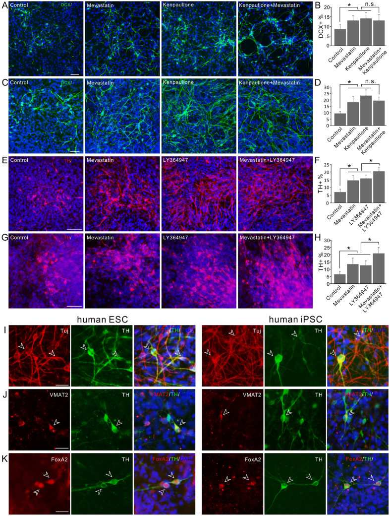
Figure 7. The effect of compounds on human PSC differentiation.
(A) Images of DCX+ neuronal cells differentiated from human ESCs. Cells were treated with 0.5 μm mevastatin; 3 μm kenpaullone; combination; or DMSO control during differentiation. (B) Quantification of the percentage of human ESC differentiated DCX+ neuronal cells with or without treatment. (C) Images of DCX+ neuronal cells differentiated from human iPSCs. Cells were treated with 0.5 μM mevastatin; 3 μM kenpaullone; combination; or DMSO control during differentiation. (D) Quantification of the percentage of human iPSC differentiated DCX+ neuronal cells with or without treatment. (E) Images of TH+ dopaminergic neuronal cells differentiated from human ESCs. Cells were treated with 0.5 μm mevastatin; 3 μm LY364947; combination; or DMSO control during differentiation. (F) Quantification of the percentage of human ESC differentiated TH+ neuronal cells with or without treatment. (G) Images of TH+ dopaminergic neuronal cells differentiated from human iPSCs. Cells were treated with 0.5 μM mevastatin; 3 μM LY364947; combination; or DMSO control during differentiation. (H) Quantification of the percentage of human iPSC differentiated TH+ neuronal cells with or without treatment. (I–K) Characterization of the TH+ cells differentiated from mevastatin LY364947 combination treated human PSCs (left panels, human ESCs; right panels, human iPSCs). All the TH+ cells expressed neuronal marker Tuj (I), most of them also coexpressed VMAT2 (J). All the TH+ cells also expressed midbrain floor plate marker FoxA2 (K). Example cells were pointed with arrow heads. *P < 0.05. Scale bars, 50 μm in (A–G); 20 μm in (I–K).

