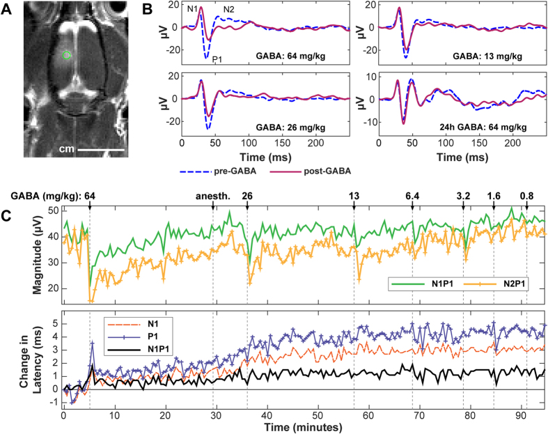
Figure 1. (a) Contrast-enhanced MRI showing ultrasound-induced BBB disruption induced in a rat’s somatosensory cortex in one hemisphere.
SSEP recordings were subsequently obtained with needle electrodes placed under the skin at the point indicated by the circle (2 mm lateral, 2 mm posterior to bregma). (b) Example SSEP recordings made in Rat 1 before and after administration of different doses of GABA. Measurements were made approximately 40–120 min after FUS-induced BBB disruption and repeated 24 hours later. (c) Changes in SSEP magnitude and latency (time-to-peak for N1, P1; time between peaks for N1P1) plotted as a function time in the same rat, who received multiple additional GABA doses over a 90 min period. Changes in latency (in ms) from that measured at the first recording are shown. The SSEP magnitude suppression and the time required to recover to pre-GABA levels were both dose-dependent. Obvious latency changes related to GABA administration were observed only at the highest dose tested. A slow increase in the time-to-peak for both N1 and P1 occurred starting at approximately 30 min. The reason for this increase is not known but may be related to administration of anesthesia a few min earlier.

