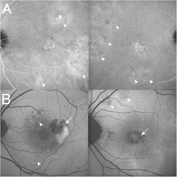
Figure 3.
(A) Choroidal vascular hyperpermeability. Choroidal vascular hyperpermeability is evidenced by focal areas of hyperfluorescence, which appear during the middle phase of indocyanine green angiography (IA) and expand over time, eventually forming a ring shape (arrowheads). The center of the initially hyperfluorescent area becomes hypofluorescent during the late phase. (B) Retinal pigment epithelium abnormality. Retinal pigment epithelium (RPE) abnormality is seen as patchy areas of granular hypoautofluorescence with occasional discrete hyperautofluorescent specks scattered throughout the fundus autofluorescence (FAF) images16,17 (arrowheads). However, this was not considered indicative of RPE abnormality when this finding was adjacent to a choroidal neovascularization (CNV; arrows) lesion.

