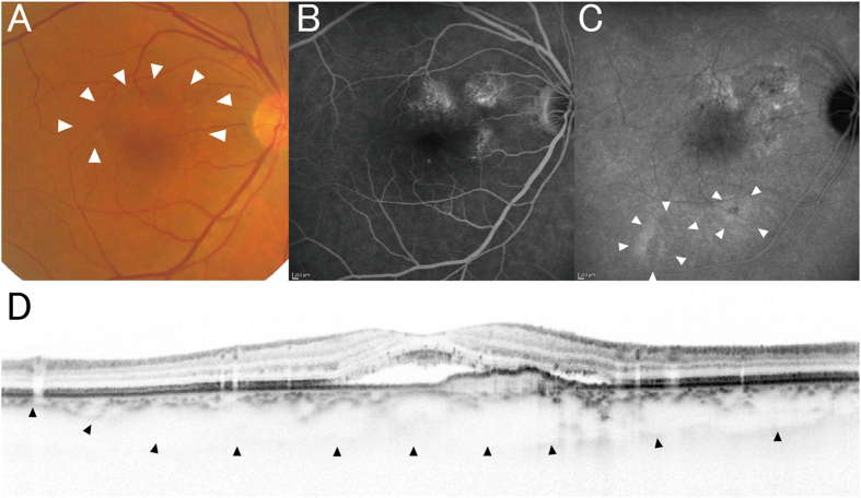
Figure 5. A case of pachychoroid neovasculopathy.
A 68-year-old male was visually impaired in the right eye (best-corrected visual acuity = 30/20). (A) Color fundus photograph shows serous retinal detachment (arrowheads) without drusen. (B) FA suggests occult CNV. (C) Late-phase IA shows choroidal vascular hyperpermeability (arrowheads). (D) Enhanced depth imaging OCT (vertical scan thorough the center of the fovea) reveals type 1 CNV and subretinal fluid. Choroid is thick throughout the macula, and subfoveal choroidal thickness was measured as 353 μm. He had GT genotype at ARMS2 A69S (rs10490924) and AG genotype at CFH I62V (rs800292).

