. 2015 Jul;96(Pt 7):1696–1701. doi: 10.1099/vir.0.000111

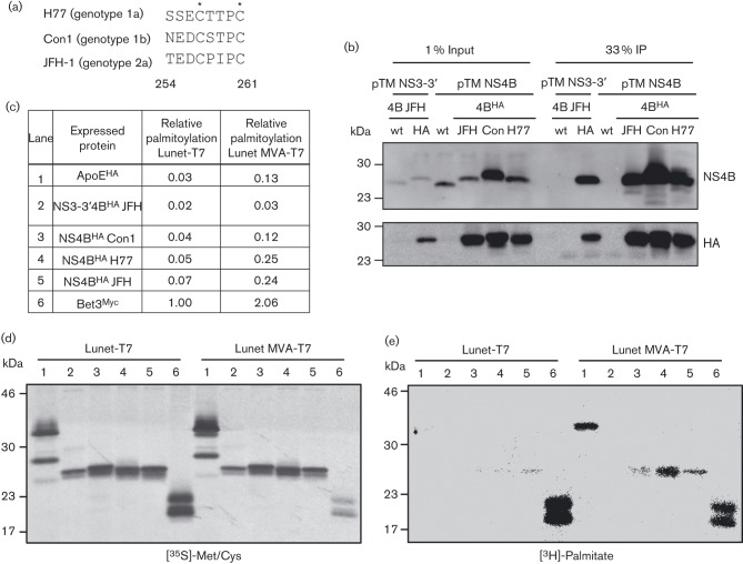
Fig. 1. The non-palmitoylated status of NS4B is altered upon MVA-T7-driven protein expression. Shown is a sequence alignment of the carboxy-terminal region of NS4B (amino acids 254–261) for the three HCV isolates used in this study, with asterisks indicating cysteine residues previously reported to be palmitoylated (a). Huh7-Lunet/T7 cells transfected with pTM expression plasmids as indicated on the top were harvested 24 h after transfection and NS4B was immunocaptured with HA-beads. Western blot analysis with mono-specific antibodies indicated on the right is shown in (b). After transfection of cells with pTM and control expression plasmids as indicated in (c), metabolic labelling with either [3H]-palmitate or [35S]-Met/Cys was performed. Proteins were purified by HA-, or Myc-specific IP and after SDS-PAGE the [35S]-labelled proteins were visualized by autoradiography (d) and the [3H]-signal was detected using a β-imager (e). A total of 33 % of the IP fraction was loaded in each lane and molecular mass standards are indicated on the left of each blot. (c) Relative palmitoylation of the respective protein was calculated as a ratio of [3H]/[35S] signal normalized for the sum of Met/Cys residues in the given protein and assuming a single palmitoylation site. Values that are normalized to the Bet3 positive control expressed in Lunet-T7 cells are shown.

Fig. 1.