. 2015 Jul;96(Pt 7):1702–1711. doi: 10.1099/vir.0.000113

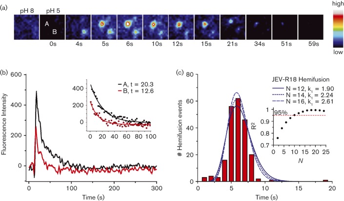
Fig. 4. TIRFM of membrane hemifusion by single JEV VLPs. (a) Snapshots (after bilinear interpolation) of two R18-labelled JEV VLPs fusing with the planar membrane. Images were false coloured with the indicated pixel value map. (b) Fluorescence intensities of the particles in (a) over the time course of fusion. Lipid mixing caused sudden dequenching of R18, followed by decay due to diffusion of the dye into the membrane. The exponential decay constant was 13–20 s (inset). (c) Distribution of time elapsed between pH drop and hemifusion of individual particles (n = 214). The best fit to a gamma distribution function was with N = 15 transitions, k 1 = 2.43 s−1. Inset, R2 values of gamma distribution fits at various N values.

Fig. 4.