. 2015 Jun;96(Pt 6):1369–1373. doi: 10.1099/vir.0.000085

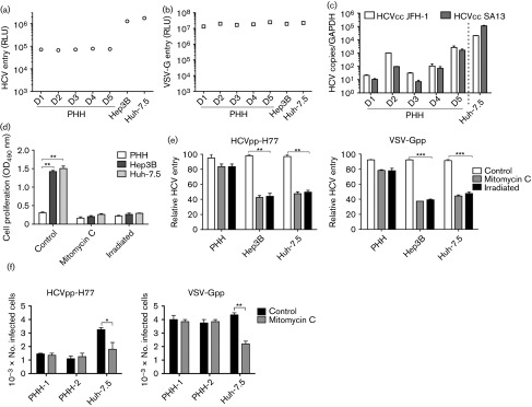
Fig. 3. Cell proliferation augments pseudoparticle infection. PHHs, and Hep3B and Huh-7.5 cells were seeded at 4×104 cm−2 on collagen-coated plates and 2 days post-plating were infected with HCVpp, VSV-Gpp (a, b) or HCV JFH-1 and SA13/JFH (c) virus for 8 h as detailed in Fig. 1. Data are presented as RLU from which a no-envelope pseudotype control value has been subtracted or as HCV RNA copy number relative to GAPDH and are representative of three independent experiments. (d) Mitomycin C and γ-irradiation block cell growth. Cells were treated with mitomycin C (10 µg ml−1, overnight) or γ-irradiated (32 mSv, 30 min) and proliferation was measured using an MTT assay (Promega) as per the manufacturer’s instructions; data are presented as OD450. (e) Mitomycin C or γ-irradiated cells were infected with HCVpp-H77 or VSV-Gpp. Cultures were maintained at 37 °C for 72 h and luciferase activity was measured as described above. Data are presented relative to untreated cells. (f) Control and mitomycin C-treated PHHs (donor 2) and Huh-7.5 cells were studied for their ability to support HCVpp-H77 and VSV-G pseudoviruses expressing green fluorescent protein (GFP) reporter. GFP-expressing cells were detected 72 h post-infection by flow cytometry. *P<0.05, **P<0.01, ***P<0.001 (t-test). Error bars indicate sd from the mean (n = 3).

Fig. 3.