. 2015 Jun;96(Pt 6):1411–1422. doi: 10.1099/vir.0.000071

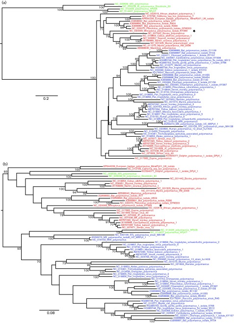
Fig. 5. Maximum-likelihood phylogenies produced from different regions of the genome. (a) Partition A, comprising the late region plus a short region of LT-Ag exon 2. (b) Partition B, comprising the majority of the early region. Red and blue coloured branches indicate the major two clades as identified in the genome-wide alignment (Fig. 1). Green branches indicate human polyomaviruses within inconsistent phylogenetic locations. The Miniopterus polyomavirus is marked with a star to indicate identification in Recco as a further possible recombinant. Scale bar represents expected number of substitutions per site (first and second codon positions only).

Fig. 5.