
Fig. 9.
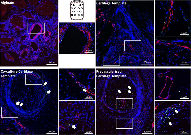
Immunohistochemical staining of the groups after 4 weeks implantation. Boxes denote area of magnification. Images were taken at 10× and 60×. Schematic of the plane in which the section was taken is in the middle of the image. CD31 stained in green, nucleus stained in blue, smooth actin stained in red. Arrows denote presence of CD31 (green) within vessel formation