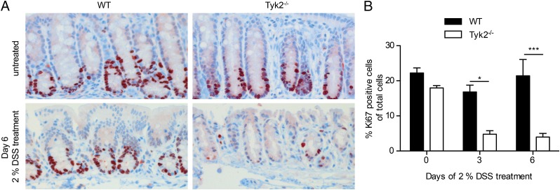
FIGURE 6.
Reduced epithelial proliferation in the absence of Tyk2. (A) Representative images of immunohistochemical staining for Ki67 on colon tissue sections of untreated and day 6 DSS-treated WT and Tyk2−/− mice. Original magnification ×200. (B) Quantification of Ki67 immunohistochemistry on WT and Tyk2−/− colon tissue sections on days 0, 3, and 6 of DSS treatment using HistoQuest software. Stainings are derived from two independent experiments with a total of six mice per genotype; a minimum of three images per mouse were quantified. Results are given as mean values ± SEM. *p < 0.05, ***p < 0.001.

