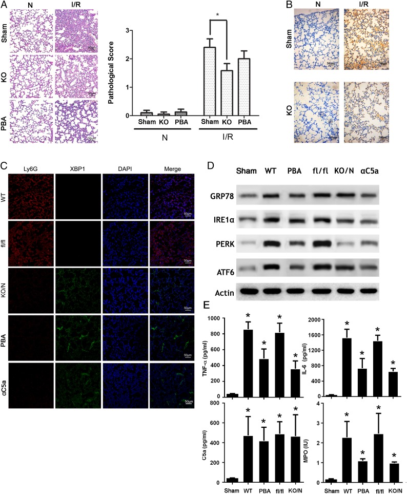
FIGURE 5.
Inhibition of ER stress in neutrophils decreases ALI in vivo. (A) xbpf/f MRP8-cre mice (KO) and XBP1f/f mice were used to establish IR-induced ALI model. Lungs samples were fixed and embedded in paraffin. Tissue blocks were sectioned at 5 μm and stained with H&E. Morphology was examined using light microscopy. Pathological scores were given by an experienced pathologist. (B) Sections were stained with Ly6G (1A8) Ab. (C) Neutrophils were prepared from the BALF of normal or ALI xbpf/f MRP8-cre mice and XBP1fl/fl mice, and they were subjected to immunofluorescence staining with indicated Abs. Images shown are representatives of three experiments. (D) Neutrophils were prepared from the BALF of normal or ALI xbpf/f MRP8-cre mice and XBP1fl/fl mice and then lysed. Lysates were analyzed by immunoblotting with indicated Abs. Images shown are representatives of three experiments. (E) BALF was collected from xbpf/f MRP8-cre mice (KO/N) and XBP1fl/fl mice (fl/fl). TNF-α, IL-6, C5a, and MPO were measured using ELISA. Quantification is expressed as mean ± SD of three independent experiments. Scale bars, 50 μm. *p < 0.05.

