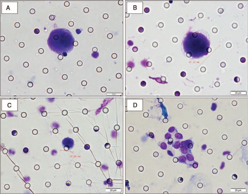
FIGURE 2.

Cytomorphological analysis of CTCs/CTM detected on filtered blood using the ISET method in patients with ESCC. (A–D) Cells showing cytological malignant features isolated by the ISET method in patients with ESCC. Romanowsky staining. (A) CTC: nuclear-cytoplasmic ratio >0.8; nucleoli is abnormally huge; (B) CTC: the diameter of the nucleus >18 μm; nuclear membranes appear thickened, sunken, wrinkled, and jagged; (C) CTC: nuclear shape is irregular; hyperchromatic nuclei with nonuniform color; presence of nuclear chromatin side shift; (D) CTM: presence of tumor cells (≥3) aggregation. The cells were analyzed under ×40 magnification. The scale bar is 20 μm.
