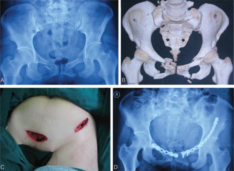
FIGURE 1.

A 28-year-old man suffered from pelvic fractures. A, The anteroposterior radiograph shows the fractures of bilateral pubic rami and sacroiliac joint of the pelvis (Type 61-B1, according to AO/OTA classification). B, Three-dimensional CT reconstruction image shows the fracture clearly and open-book dislocation of sacroiliac joint. C, It shows the minimally invasive ilioinguinal approach, which was composed of the lateral and the medial portions of the standard ilioinguinal approach. D, Postoperative anteroposterior radiograph after the internal fixation of the pelvic fracture shows the good quality of fracture reduction.
