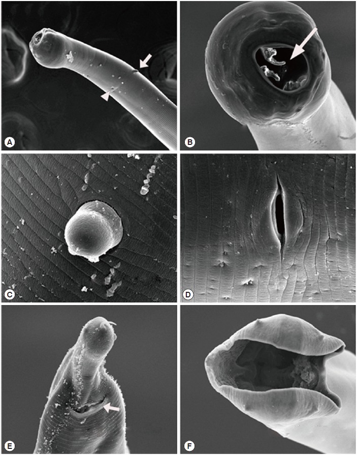
Fig. 2.

Scanning electron micrographs of Globocephalus samoensis isolated from the small intestine of a Korean wild boar (Sus scrofa coreanus) from South Korea. (A) Anterior end of a female, illustrating 1 of the 2 cervical papillae (white arrow) and an excretory pore (white arrow head). (B) Enlarged enface view of the anterior end showing the 2 bicuspid lancets (white arrow). (C) Enlarged view of the cervical papillae. (D) Enlarged view of the vulva. (E) Posterior end of female, showing the anus (white arrow). (F) Enface posterior end of a male, showing the copulatory bursa.
