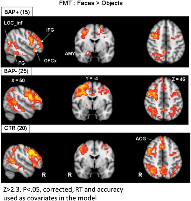
Figure 5.
Voxel-based whole-brain analysis during the FMT. Highlighted here are areas of increased activation (faces > objects) along broad regions of temporal and occipital cortex, as well as the amygdala in all subject groups. AMY, amygdala; FG, fusiform gyrus; OFC, orbital frontal cortex; IFG, inferior frontal gyrus; LOC_inf, inferior lateral occipital cortex; ACG, anterior cingulate gyrus; RT, reaction time; ACC, accuracy. Areas of activation passed a cluster-significance threshold of z > 2.3, with whole-brain cluster-correction at P ≤ 0.05. R indicates right.

