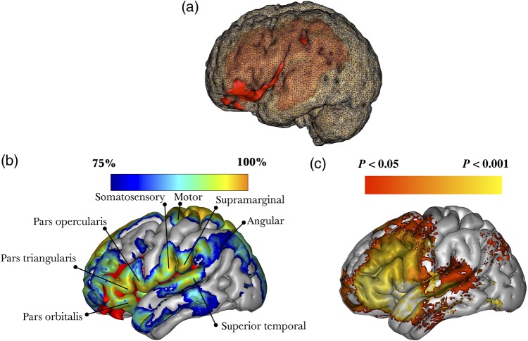
Figure 7.
3D reconstruction of Leborgne's brain in MNI space. (a) The lesion (red) damaged both cortical structures (posterior inferior frontal cortex) and subcortical white matter (perisylvian pathways). (b) 3D reconstruction of the MNI152 template, with a blue-to-orange gradient indicating the probability of disconnection of those areas not directly affected by the lesion; red color indicate damage caused by the lesion. (c) Meta-analysis of functional MRI studies reporting activations related to the performance of fluency tasks (75 studies).

