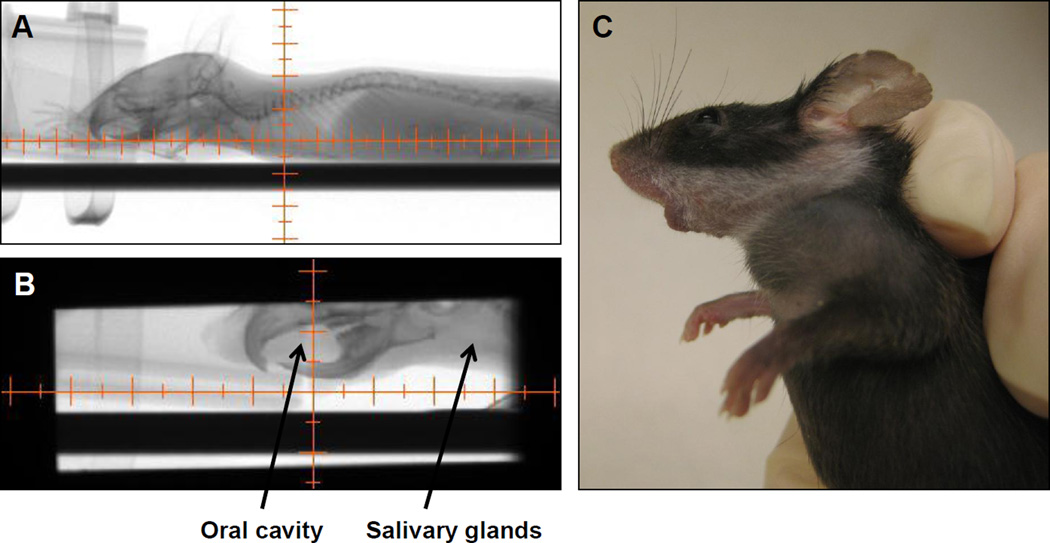
Figure 1.
Irradiation using the X-RAD 225Cx small animal irradiator. A: Mice were visualized using the fluoroscopy settings. B: The collimator’s isocenter was placed in a position that targeted radiation beam to include the oral cavity and salivary glands while excluding the eyes and central nervous system. C: The resulting leukotrichia post-RT, reflecting the precision of the radiation beam.

