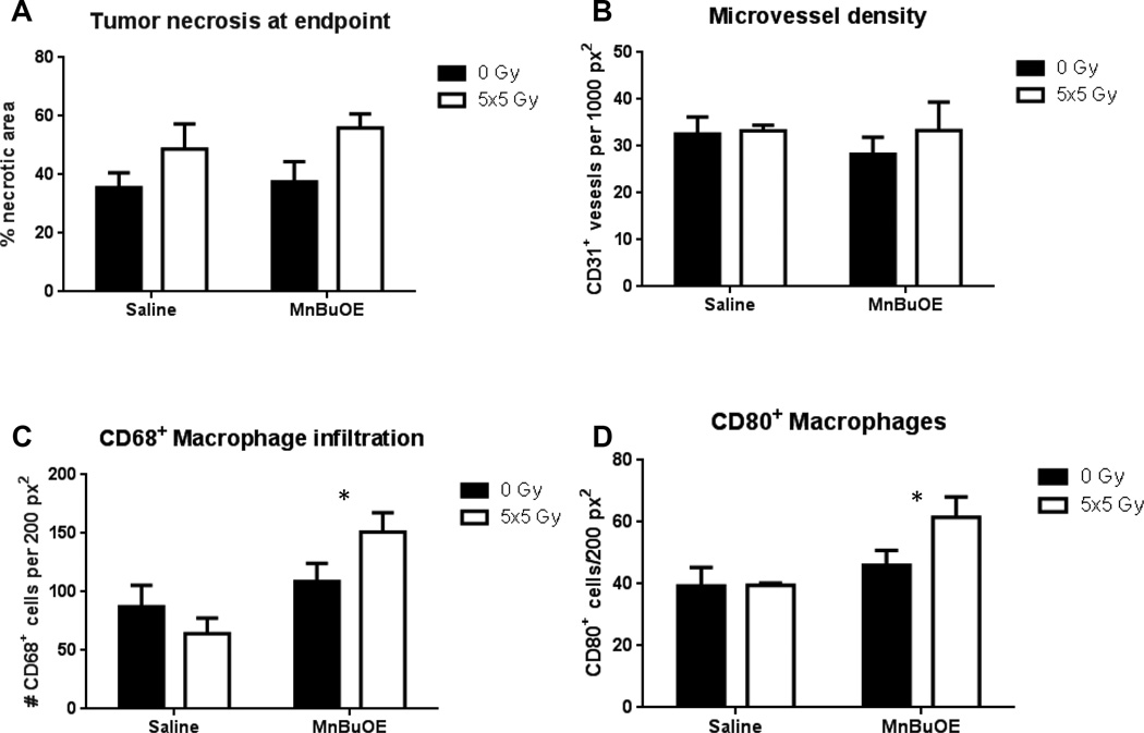
Figure 6.
Immunohistochemistry of tumors taken at endpoint (volume >1500 mm3). A: Percentage of tumor that was necrotic. B: Microvessel density. C: Macrophage infiltration, using CD68 as a pan-macrophage surface marker. p=0.01 D: M1 macrophage infiltration, using CD80 as the identifying surface marker p=0.03. N=3–6/group. Error bars represent the standard error.

