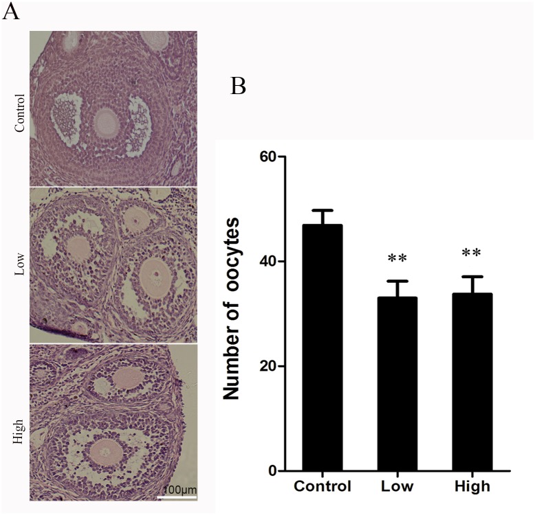
Fig 4. Effect of melamine on ovarian and histological analysis of the ovary.
Hematoxylin and eosin staining was performed on paraffin sections of mouse ovary. The follicles showed normal cell associations with a lot of granulosa cells in the control group. The granulosa cells apoptosis and disrupted structure in the treatment groups. Data show the means ± SE from at least three separate experiments. **P <0.01 versus control.

