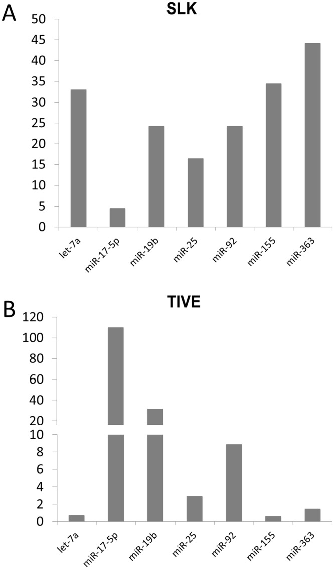
Fig 1. Quantitative RT-PCR analysis of miRNAs in latently KSHV-infected SLK and TIVE cells.

The expression differences of miRNAs in KSHV-infected SLK (A) and TIVE cells (B) from non-infected SLK and TIVE cells. U6 expression was used as internal control for normalization.
