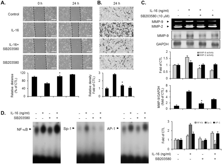
Fig 4. Inhibition of p38MAPK interfered with the IL-16-stimulated induction of migration, invasion, MMP-9 expression, and the Sp-1 motif activation in VSMCs.
Quiescent VSMCs were pre-cultured with SB203580 (10 μM) for 40 min, prior to IL-16 (50 ng/ml) stimulation for 24 h. (A, B) Indicated cells were examined to determine the migratory capacity and invasive potential using wound-healing migration and matrigel invasion assay. Scale bars represent 400 μm (wound-healing) and 100 μm (invasion). *P < 0.01 compared with IL-16 treatment. (C) Production of MMP-9 was analyzed by gelatin zymography using the conditional medium from indicated cells. Cell lysates were examined to determine the protein level of MMP-9 via immunoblot experiment. *P < 0.01 compared with IL-16 treatment. (D) EMSA was performed with nuclear extract from indicated cells to determine the activation of NF-κB, AP-1, and Sp-1 binding motifs using radiolabeled oligonucleotide probes. Results are reported as the means ± SE from three triplicate experiments. *P < 0.01 compared with IL-16 treatment.

