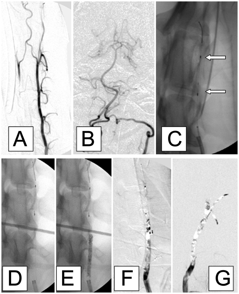
Fig 1. Representative angiographic images from a dog showing the successful establishment of left vertebral artery occlusion.
A, B: Antero-posterior images of the left vertebral artery after injection of contrast. C, D: A custom-designed, dedicated, self-expanding thrombus filter (length 10 mm, diameter 4 mm; white arrows, panel C) was pre-deployed into the middle segment of the left vertebral artery. E: The non-subtracted image shows adequate radiopacity of the injected barium sulfate-marked thrombus. F: Digital subtraction image showing complete occlusion of the left vertebral artery. G: Complete occlusion of the vessel was confirmed 45 min later. Vertebrobasilar embolism did not occur distal to the thrombus filter.

