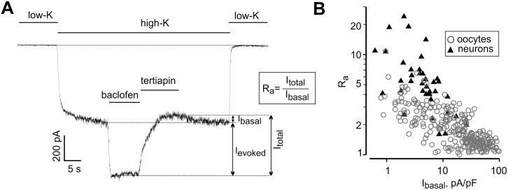
Fig 1. Basal and agonist-evoked GIRK currents in neurons and oocytes are inversely related.
(A) A representative whole-recording of GIRK current in a neuron. Switching from low-K+ extracellular solution to a high-K+ solution led to the development of a large inward current probably carried by several ion channel types. Addition of baclofen elicited Ievoked. Arrows show the amplitudes of Ibasal, Ievoked and Itotal. Extent of activation, Ra, is defined as Itotal/Ibasal. (B) Inverse correlation between Ibasal and Ra in oocytes and neurons. To allow direct comparison of Ibasal in oocytes and neurons, currents in neurons were corrected for the 10 mV difference in holding potential, which was -70 mV in neurons and -80 mV in oocytes (see Methods). The correlation between Ra and Ibasal was highly significant, p = 0.000000028 (neurons; n = 60; correlation coefficient = -0.633) and p = 0.0000002 (oocytes; n = 272; correlation coefficient = -0.728) by Spearman correlation test.

