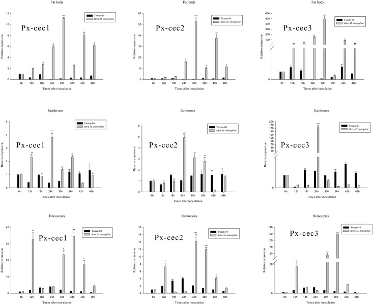
Fig 2. qRT-PCR analysis of relative expression of cecropin genes from fat body, epidermis, hemocytes after inoculation of M. anisopliae.
Actin was used as an internal control. The mRNA levels of cecropins were highly expressed in 30 h after induction of M. anisopliae in fat body and 24 h in epidermis, Px-cec1, Px-cec2 and Px-cec3 showed maximum expression after 36 h, 30 h, 36 h in hemocytes, respectively. In addition, Px-cec3 depicted more sensitivity to M. anisopliae than Px-cec1 and Px-cec2. Relative expression levels of 6 h was arbitrarily set at 1. Three biological replications (n = 3) were conducted, and the 2-ΔΔCt method was used to measure the relative transcription levels. Means with different number of asterisk are significantly different (P<0.05) (Duncan’s Multiple Range Test) among different time after treated with alive M. anisopliae. *: different with the lowest expression after treated with M. anisopliae; **: significant different with the lowest expression after treated with M. anisopliae; ***: highly significant different with the lowest expression after treated with M. anisopliae.

