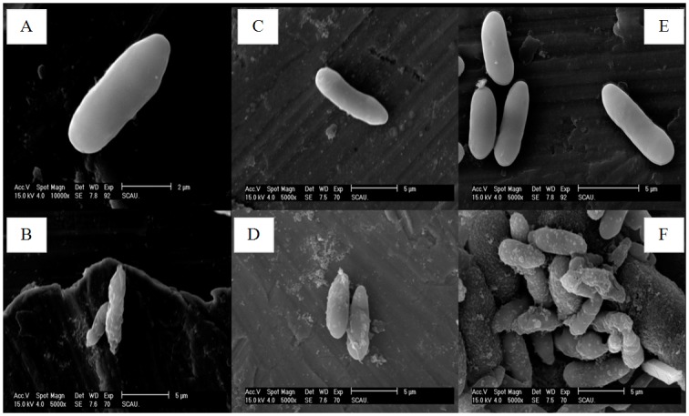
Fig 7. SEM analysis of the spore of M. anisopliae interacted with cecropins.

A, C and E: CK (naive M. anisopliae spore); B, D and F: the spore of M. anisopliae interacted with Px-cec1, Px-cec2 and Px-cec3, respectively. The SEM analysis showed that M.anisopliae spore became short and wrinkled (B, D and F) after interacted with cecropins from P. xylostella as compared to the untreated spore (A, C and E), which had a bright and normal smooth surface.
