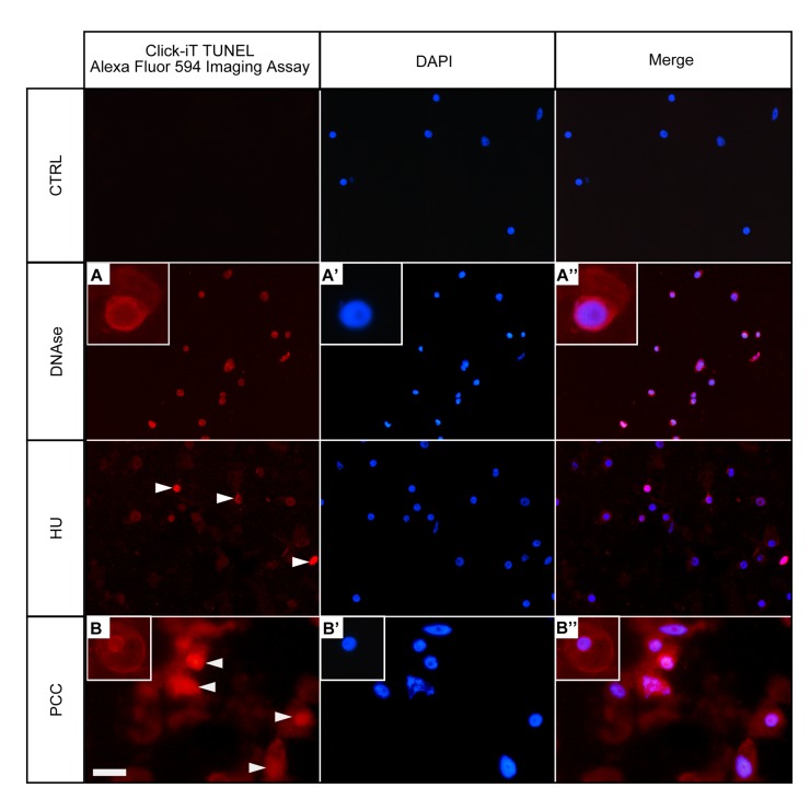
Fig 3. Terminal deoxynucleotidyl-dUTP nick end labeling (TUNEL) assay in Click-iT technology in the untreated control (negative), DNase-treated control (positive), HU-treated and HU/CF-co-treated (i.e. PCC-induced) Vicia faba root meristem cells.
Left panel—DNA fragmentation in V. faba cells detected by TUNEL reaction and visualized by AlexaFluor 594. Central panel—DAPI stained nuclei. Right panel—Merged images (AlexaFluor 594 + DAPI). Positively stained nuclei appear in the DNase-treated cells (e.g. A-A'') and in the HU/CF co-treated cells (e.g. B-B''). Positively stained nuclei in the HU-treated series are indicated by arrowheads. Non-reacting nuclei can be seen in the negative control sections (as indicated in the highest panel described as 'CTRL'). Scale bar = 20 μm.

