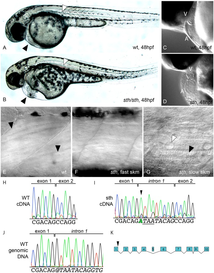
Fig 1. Still heart, a smyd1b mutant, has defects in heart and fast skeletal muscle tissue.
A lateral view of 48hpf wild type (A) and still heart (sth) mutants (B), which have pericardial edema, small eyes, malformed head and reduced motility. Black arrowheads highlight the pericardial edema in sth mutants and the absence of edema in wild type. White arrowheads indicate blood pooling in the mutant and the absence of pooling in wild type. (C&D) Sth mutant hearts are underdeveloped and do not beat. (E-G) Examination of lateral myofibers at 5dpf under DIC microscopy revealed striations, indicative of fully formed sarcomeres, are visible in the myofibers of wild type muscle (E, black arrowheads), while absent in the fast muscle of still heart mutants (F); striations are present in sth slow muscle (G, black arrowhead) but are disturbed by nuclei and fluid-filled spaces (G, white arrowhead). Sequencing of smyd1b cDNA from wild type embryos (H) and sth mutant embryos (I) revealed a 9 nucleotide insertion between exon 1 and 2 in the smyd1b mRNA, creating an in-frame stop codon (I, underlined sequence). The insertion is the first 9 nucleotides of intron one as sequenced from wild type smyd1b genomic sequence (J). This is a result of a transition mutation in the splice donor site of intron 1 (I, green letter in sequence, J, outlined letter in sequence). This results in a premature truncation of the SMYD1b protein after exon 1 (K).

