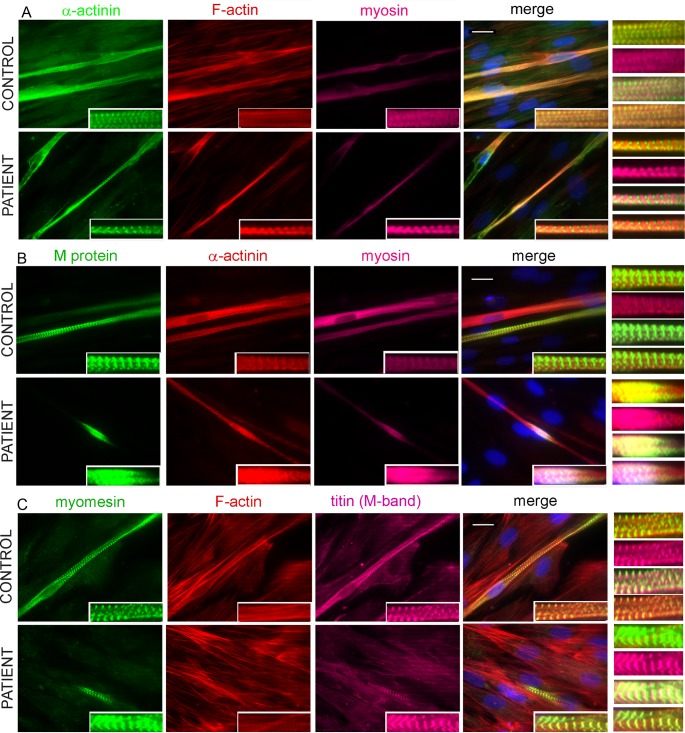
Fig 4. Formation of sarcomere structure of 6-day differentiated myotubes.
(A) Triple staining was performed for α-actinin (green), F-actin (red), and myosin (magenta); (B) M-protein (green), F-actin (red), and myosin (magenta); (C) myomesin (green), F-actin (red), and M-band epitope of titin (magenta). The results were visualized with a Zeiss Axio Observer microscope (Carl Zeiss AG, Germany) at 63× magnification. All nuclei were counterstained with DAPI (blue). The repetitive well-structured sarcomere can be seen clearly in control myotubes and patient myotubes (insets). The bars represent 10 μm.

