
Fig 4. Model CPMG relaxation rate as a function of inter-echo time τ 180.
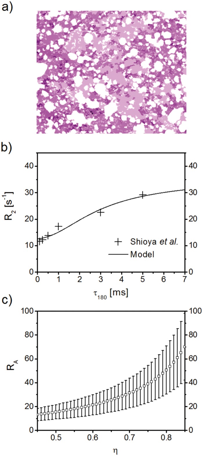
(a) Sketch of passively deflated lung tissue, modified from [23]. Air filled spaces or alveoli for passively deflated lung tissue are less numerous and prominent than in non-deflated lung tissue. (b) Relaxation rate R 2 for passively deflated lung tissue (continuous line) in comparison with experimental data [23]. The analytical model is fitted to the experimental data points, with resulting fitted values of characteristic time τ = 0.56 ± 0.22 s (p = 0.088) and intrinsic relaxation rate R 2,0 = 12.58 ± 0.96 s−1 (p = 9.72 ⋅ 10−4). With the use of Eq (5), the mean alveolar radius follows as R A = 31.46 ± 13.15 μm, which is in very good agreement with the expected value of ∼34 μm [41, 51]. (c) Model mean alveolar radius R A for different air volume fractions η (error bars represent the standard error of R A from the model fit; p-values never exceeded 0.088). Naturally, the mean alveolar radius increases with increasing air volume fraction and reaches a value of R A = 70.12 ± 28.04 μm for η = 0.85.