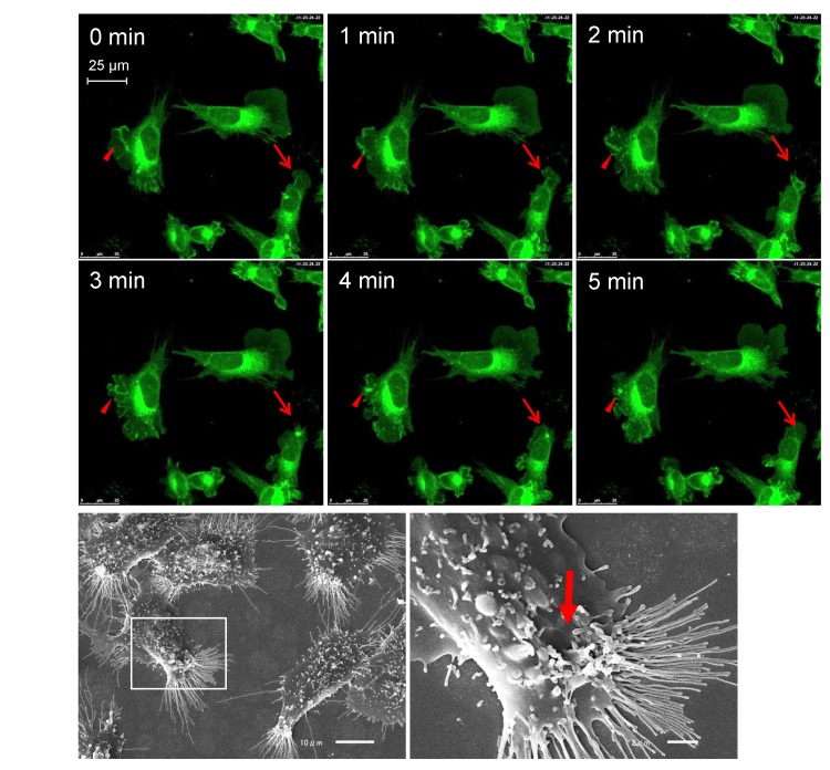
Fig 1. Macropinocytic activity of MARCO-GFP CHO cells.
(A) The cells were grown in glass-bottom culture dishes in F12 complete medium and observed using confocal microscopy. Fluorescence images were recorded at 1-min intervals. The arrows and arrowheads indicate the formation and fate of two separate macropinocytic sites. See also S1 Movie. (B) Scanning electron micrographs of GFP-MARCO-CHO cells. The right panel provides a higher magnification image of the boxed area indicated in the left panel. The arrow indicates the macropinocytic structure.

