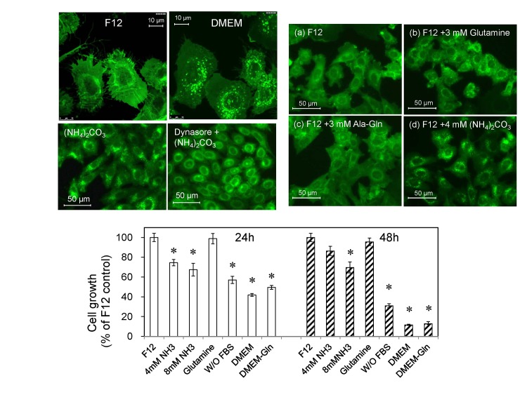
Fig 2. L-Glutamine and ammonia induce fluorescent puncta in GFP-MARCO-CHO cells.
(A) The cells were cultured in either F12 complete medium or DMEM complete medium and the fluorescence images were captured by confocal microscopy. Many puncta (200–600 nm in diameter) appeared when the cells were culture in DMEM. (B) The cells were cultured for 12 hr in (a) F12 complete medium (containing 1 mM L-glutamine), (b) F12 medium supplemented with 3 mM L-glutamine (to a final concentration of 4 mM L-glutamine), (c) F12 medium supplemented with 3 mM L-alanyl-L-glutamine (Ala-Gln), or (d) F12 medium supplemented with 4 mM (NH4)2CO3. (C) Proliferation of GFP-MARCO-CHO cells in different culture media. The cells were suspended in F12 medium at 2.0 x 10⁴ cells /mL, aliquotted at 100 μL/well in a 96-well culture dish, and pre-cultured overnight. The medium then was replaced with fresh medium as follows: F12 medium; F12 medium supplemented with 4 or 8 mM (NH4)2CO3; F12 containing 4 mM L-glutamine, F12 without 10% FBS; DMEM; or L-glutamine-free DMEM. Cells then were cultured for 24 h or 48 h. The viable cells were assayed using WST-8. Data are presented as mean ± SEM of 6 wells per medium per time point. *, Significantly different from control (F12) value. (D) Loss of ammonia-induced fluorescent small puncta upon inhibition of the endocytic pathway. The cells were cultured in F12 culture medium and exposed to 4 mM ammonium carbonate for 6 hr in the absence or presence of 100 μM dynasore, a dynamin inhibitor.

