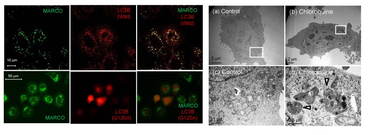
Fig 3. Effects of chloroquine on fluorescent puncta formation in GFP-MARCO-CHO cells.
(A) Co-localization of green fluorescent puncta with LC3B. GFP-MARCO-CHO cells were transfected with constructs encoding LC3B (wild-type or G120A mutant), pre-cultured overnight in F12 complete medium, and further cultured for 16 hr in F12 complete medium supplemented with 50 μM chloroquine. (B) Transmission electron micrographs of GFP- MARCO-CHO cells. The cells were cultured for 6 hr with (b, d) or without (a, c) 50 μM chloroquine. Fig 3B(c) and (d) provide higher magnification images of the boxed areas in (a) and (b), respectively. The arrowheads indicate autophagosomes.

