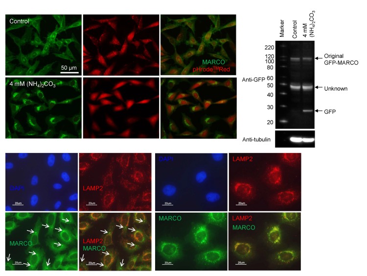
Fig 4. Processing of GFP-MARCO in lysosomes.
(A) GFP-MARCO-CHO cells were incubated with or without (NH4)2CO3 and the cells were loaded with pHrode Red AM. The cells were washed and cultured in HEPES-buffered HBSS for 2h. The acidity of lysosomes was neutralized by (NH4)2CO3. (B) Macropinocytic inclusions (arrows) did not co-localize with lysosomes. The control GFP-MARCO-CHO cells were fixed and stained with anti-LAMP2 antibody (2nd Ab: Alexa Fluor® 594-labeled goat anti-mouse IgG). (C) Co-localization of GFP-MARCO and LAMP2 in autophagic puncta. The cells were grown for 15 hr in the presence of 50 μM chloroquine. See also the legend to Fig 4 (B) for immunofluorescent staining. (D) GFP-MARCO-CHO cells were cultured for 15 hr in fresh F12 medium in the presence or absence (Control) of 4 mM (NH4)2CO3. The supernatant of the cellular lysate was analyzed by SDS-PAGE followed by western blotting for the detection of GFP. The membrane was reprobed with POD-tagged anti-tubulin to serve loading control.

