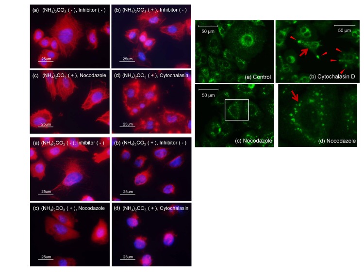
Fig 5. Effects of cytochalasin D and nocodazole on fluorescent autophagic puncta in GFP-MARCO-CHO cells.
(A) Fluorescence staining of actin in GFP-MARCO-CHO cells. The cells were cultured for 15 hr in the absence (a) or presence of 4 mM (NH4)3CO3 (b-d), as follows: (b) without further stimulation; (c) with 1 μM nocodazole; or (d) with 1 μg/mL cytochalasin D. They were fixed with formalin solution and stained with rhodamine phalloidin and DAPI using standard immunofluorescence techniques. (B) Fluorescence staining of tubulin in GFP-MARCO-CHO cells. The cells were cultured and treated with chemicals as shown in Fig 5(A). They were fixed with formalin solution and treated with anti-α-tubulin followed by Alexa Fluor® 594-conjugated secondary antibody and DAPI using standard immunofluorescence techniques. (C) The cells were cultured in the presence of 4 mM (NH4)3CO3 for 15 hr as follows; (a) without further stimulation, (b) with 1 μg/mL cytochalasin D, or (c) with 1 μM nocodazole. Fig 5C(d) provides a higher magnification image of the boxed area in (c). The arrows and arrowheads indicate autophagic puncta and interrupted macropinocytosis, respectively.

