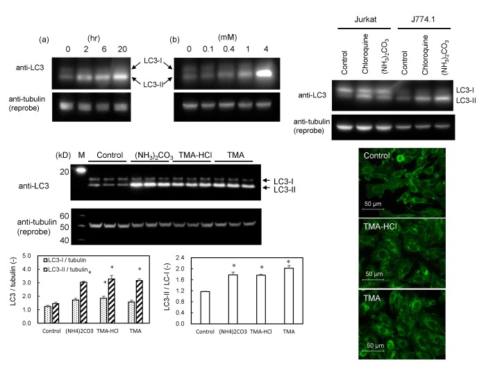
Fig 6. Effects of ammonia and amine compounds on conversion of LC3-I to LC3-II (lipidated LC3-I) and autophagic puncta formation.
(A) Western blot analysis for the detection of LC3-I and LC3-II. GFP-MARCO-CHO cells were cultured in F12 culture medium and exposed to (NH4)3CO3. (a) The cells were cultured in the presence of 4 mM (NH4)2CO3 for 0, 2, 6, and 20 hr. (b) The cells were cultured for 6 hr in the presence of 0, 0.1, 0.4, 1, and 4 mM (NH4)2CO3. α-Tubulin was adopted as loading control. The band for LC3-I was not clear in GFP-MARCO-CHO cells. (B) Jurkat (human T cell leukemia) and J774.1 (murine macrophages) cells were exposed for 6 hr to 4 mM (NH4)2CO3 or 50 μM chloroquine. (C) GFP-MARCO-CHO cells were grown for 8 hr in the absence (Control) or presence of 4 mM (NH4)2CO3, 8 mM trimethylamine hydrochloride (TMA-HCl), or 8 mM trimethylamine (TMA). M, Western marker lane. α-Tubulin was adopted as loading control and the amount of LC3-I, LC3-II and tubulin were measured by densitometry. LC3-I/tubulin, LC3-II/tubulin, and LC3-II/LC3-I ratios were presented as mean ± SEM (N = 3). *, Significantly diferent from the control value. (D) Formation of fluorescent autophagic puncta by amines. GFP-MARCO-CHO cells were cultured for 8 hr in complete F12 culture medium in the absence (Control) or presence of 8 mM trimethylamine hydrochloride (TMA-HCl) or of 8 mM trimethylamine (TMA).

