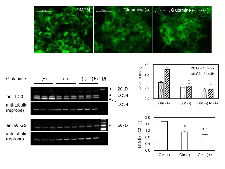
Fig 7. Autophagy in GFP-MARCO-HEK cells is induced by L-glutamine and amines.
(A) The cells were cultured (a) in DMEM complete medium for 6 hr; (b) in L-glutamine-free DMEM for 6 hr; or (c) in L-glutamine-free DMEM for 6 hr and then in DMEM (4 mM L-glutamine) for 4 hr. Fluorescent puncta appeared at 4 hr in the presence of 4 mM L-glutamine. The images were captured by fluorescence microscopy. (B) Western blot analysis of LC3 and ATG5 in GFP-MARCO-HEK cells. The cells were cultured in complete DMEM (4 mM L-glutamine) (left three lanes), L-glutamine-free DMEM for 6 hr (middle three lanes), or L-glutamine-free DMEM for 6 hr followed by culturing in complete DMEM (4 mM L-glutamine) (right three lanes). M, Western marker lane. α-Tubulin was adopted as loading control and the amount of LC3-I, LC3-II and tubulin were measured by densitometry. LC3-I/tubulin, LC3-II/tubulin, and LC3-II/LC3-I ratios were presented as mean ± SEM (N = 3). *, Significantly different from glutamine (+). #, Significantly different from glutamine (-).

