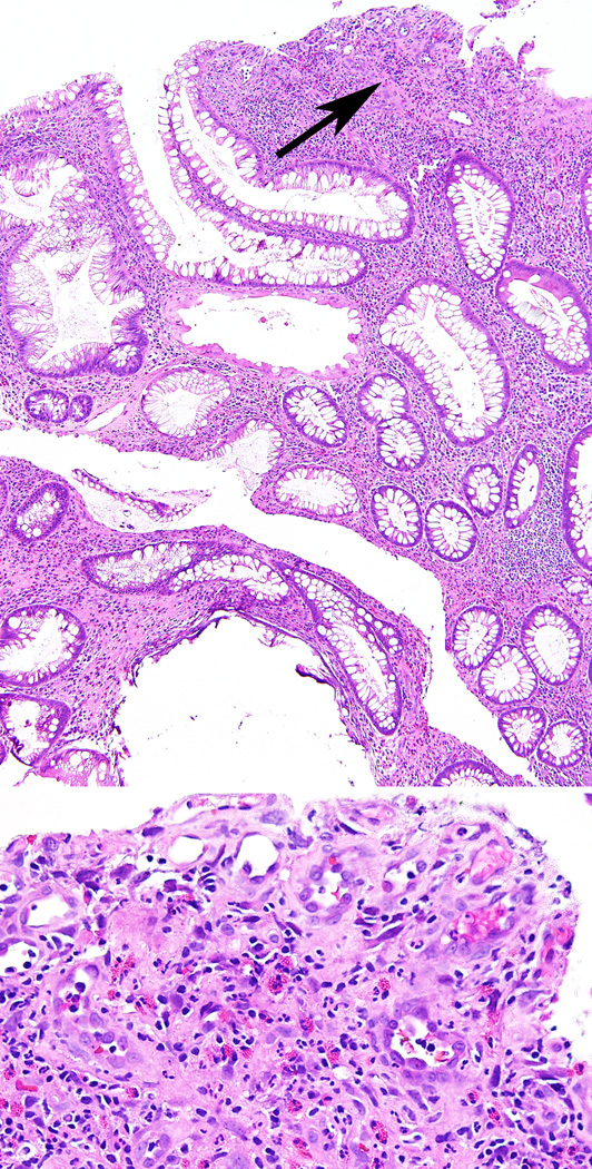
Figure 2.
Microphotograph of a small colonic juvenile polyp from the patient with NF1 and JPS showing expansion of the lamina propria with abundant inflammatory infiltrate and cystically dilated colonic crypts. In addition, there is surface erosion, granulation tissue and ‘atypical’ stromal cells, mainly representing reactive endothelial cells (arrow top panel and magnified in bottom panel).

