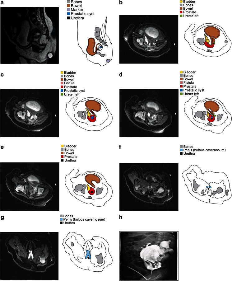
Fig. 1.
a–h A 2-month-old boy with proven recto-bladder neck fistula. a represents a midsagittal view through the pelvis (T2-weighted fast spin echo sequence; slice thickness 1.5 mm) with s-form of the distal rectal segment which enters the prostate from posterior. The axial MRI slices (b–g) (T2-weigthed fat-suppressed fast spin echo sequence; slice thickness 1.5 mm) are shown from higher to lower levels in the pelvis. The rectum turns in a fistula (pink) with a short transprostatic course. This fistula ends in the bladder neck (yellow), which turns in the urethra (black). Although on first sight of a complex case, all elements could easily be discerned by both readers based on a combination of axial and sagittal views. On the other hand, correct analysis of colostography (h) images was found to be impossible, mainly due to overlapping contrast opacities in all directions

