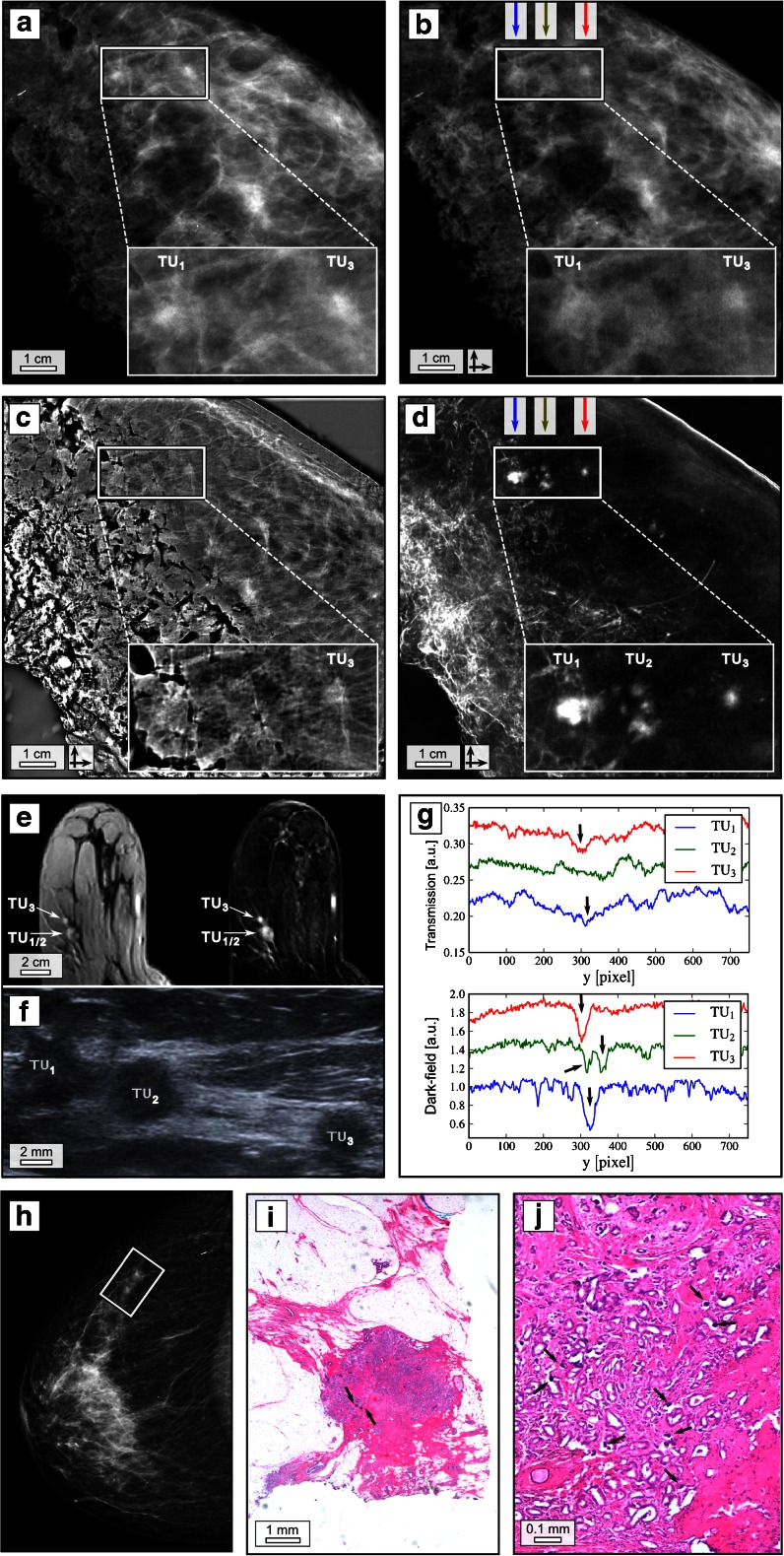
Fig. 1.
Dark-field mammography reveals radiographically undetectable tumour nodules. Clinical ex vivo mammography in craniocaudal (cc) projection at 31 kVp, 45 mAs and 0.56 mGy average glandular dose (silver filter) (a), experimental absorption-contrast mammography (abs-Mx, b), phase-contrast mammography (PC-Mx, c) and dark-field mammography (DF-Mx, d) of patient 1 at 40 kVp, 70 mA and 66 mGy mean glandular dose (per scan direction); representative section from the in vivo MRI (e) including contrast-enhanced T1 weighted gradient-echo sequence after manual injection of 0.2 ml/kg meglumine gadopentetate (Magnograf ® 0.5 mmol/ml) (e, left) and the corresponding first subtraction image after 2 min (e, right) in an axial view using a dedicated sensitivity-encoding-enabled bilateral breast coil with a 3.0-Tesla system (voxel size 0.7 × 0.7 × 2.0 mm), field of view 360 mm; base resolution 512; flip angle 15°; repetition time 6.5 ms; echo time 2.47 ms. Ultrasound (f); vertical line plots quantifying superior depiction of tumor (TU) 1, 2 and 3 in DF-Mx (g, bottom) in comparison with abs-Mx (g, top) as indicated by arrows; in vivo mammography of patient 1 in cc projection at 29 kVp, 11 mAs and 0.41 mGy average glandular dose (aluminium filter) (h); exemplary histological image (haematoxylin–eosin staining) of one calcified tumour nodule (i and j), arrows indicating microcalcifications. The rectangles in a–d and h indicate TU 1–3. The crossed arrows in b–d indicate bidirectional measurements

