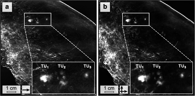
Fig. 4.
Comparison of low- and high-dose dark-field mammography. Low-dose (22 mGy mean glandular dose) dark-field mammography of patient 1 conducted in one scan direction and 3 s exposure time per phase step (a) and corresponding high-dose measurements (66 mGy mean glandular dose per scan direction) conducted in two scan directions and 9 s exposure time per phase step (b) at 40 kVp/70 mA, respectively. Both images offer equal quality in the detection of the tumour nodules

