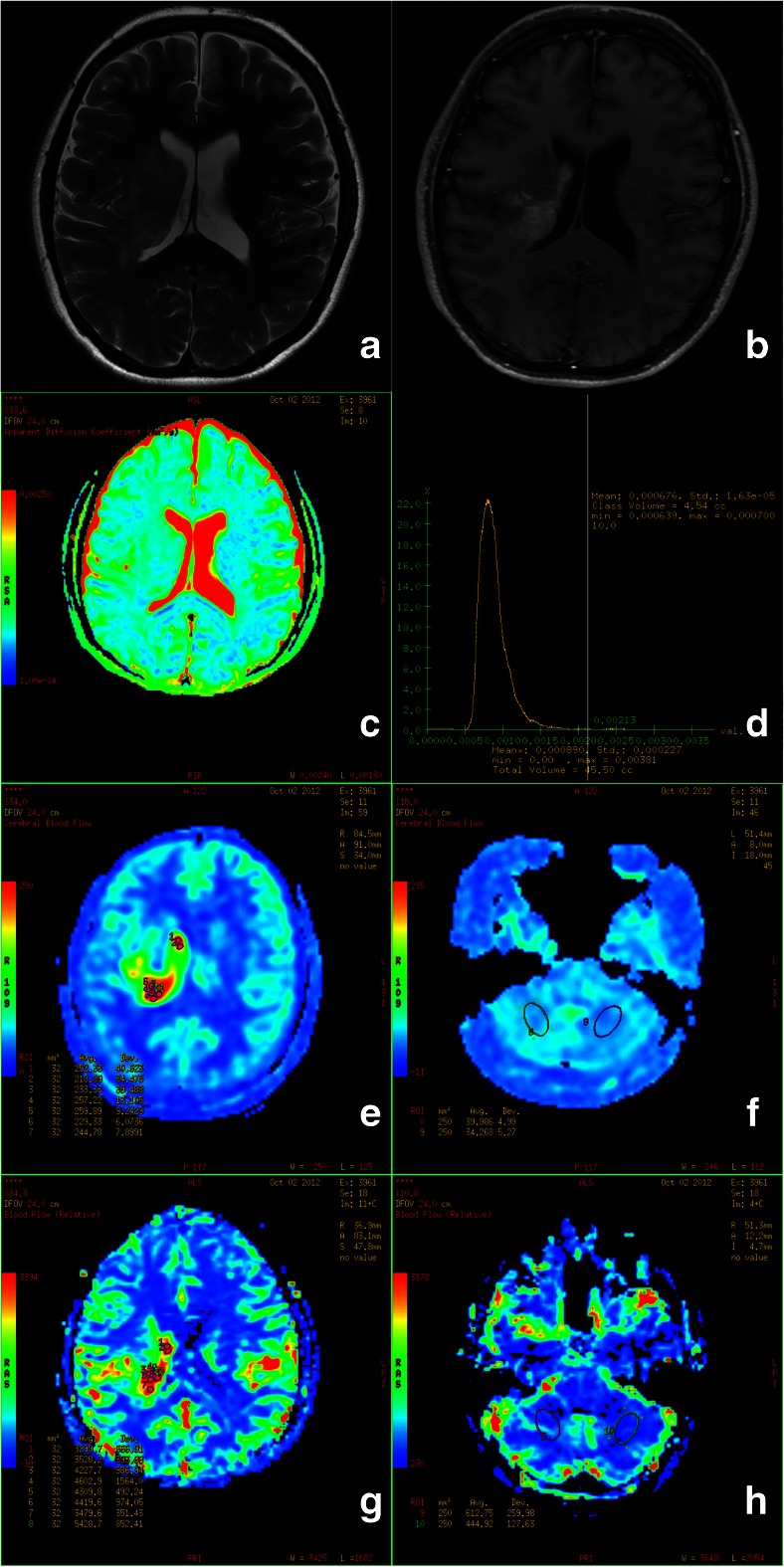
Fig. 1.
A sixteen-year-old girl with anaplastic astrocytoma (WHO III) in the right basal ganglion and thalamus. a The tumour showed hyperintense signal on T2WI. b The tumour showed patchy enhancement on post-contrast T1WI. c ADC map. d ADC histogram of the tumour. e ROI selection for the maximal CBF of the tumour on pCASL CBF color map. f ROI selection for bilateral cerebellum CBF on pCASL CBF color map. g ROI selection for the maximal rCBF of the tumour on DSC-PWI rCBF color map. h ROI selection for bilateral cerebellum rCBF on DSC-PWI rCBF color map

