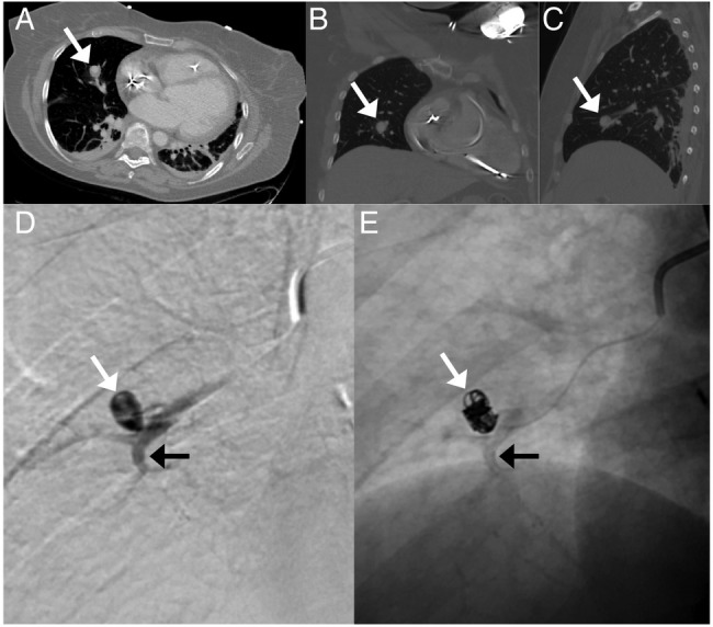
Figure 1.

CT and digital subtraction angiography (DSA) images of the pulmonary artery pseudoaneurysm. Axial (A), coronal (B) and sagittal (C) views of the pseudoaneurysm in the right middle lobe (white arrow). (D) DSA following contrast injection from the microcatheter inside the pseudoaneurysm (white arrow) arising from a right middle lobe medial segment artery (black arrow). (E) Subsequent coil embolisation of the pseudoaneurysm (white arrow); flow to the distal pulmonary artery (black arrow) is preserved.
