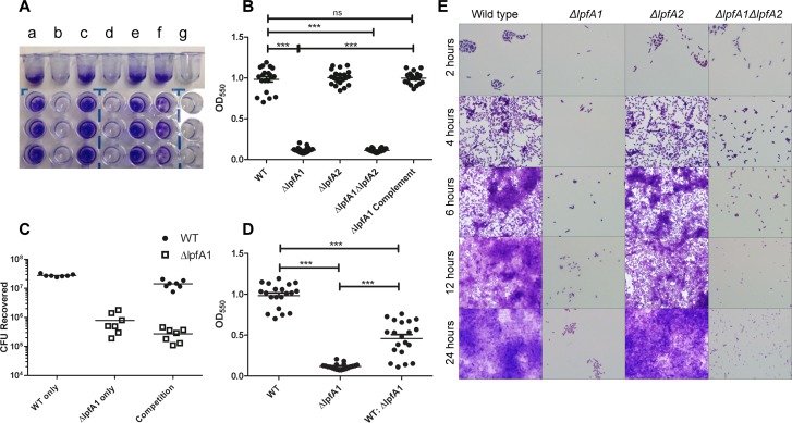
Fig 4. ΔlpfA1 and ΔlpfA2’s effect on stable biofilm formation.
Microtiter plates were washed and stained with crystal violet and biofilms visualized in (A) and quantified in (B) of E. coli O104:H4 (a), ΔlpfA1 (b), ΔlpfA2 (c), ΔlpfA1 ΔlpfA2 (d), ΔlpfA1 complemented (e), O104:H4 wt and ΔlpfA1 mix (f) or MEM media alone (g) after 24 h of static incubation at 37°C. Competition assays were done with O104:H4 2011 c3494 wild type and ΔlpfA1 mutant, and results are expressed as absorbance at 550 nm (C) and CFUs recovered (D). Formation of biofilm by all E. coli O104:H4 wild type and mutant strains were visualized by growing on glass cover slips, stained and imaged at 2, 4, 6, 12, and 24 h post-infection (E). All experiments were done with a 1:100 dilution of overnight cultures diluted to and OD600 of 1.0. Error bars indicated SEM of 3 independent experiments each performed with triplicate wells.

