
Figure 1. Hypocellularity and CD4+ CD8+ double positive (DP) lymphocyte subset depletion in the thymi of septic mice.
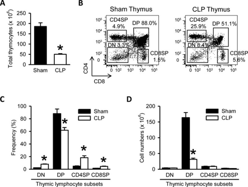
(A) Total numbers of thymocytes isolated from the thymi harvested after 20 h from sham- or CLP-operated mice are shown. (B) Flow cytometric analysis of surface CD4 and CD8 expression on the gated live lymphocytes isolated from the sham and CLP thymi. Numbers adjacent to the outlined areas in the representative dot plots show the percentage of various thymic lymphocyte subsets as indicated. DP indicates CD4+CD8+ double positive thymocytes, DN indicates CD4−CD8− double negative thymocytes, CD4SP indicates CD4+ single positive thymocytes, and CD8SP indicates CD8+ single positive thymocytes. Data are representative of three independent analyses with a total of four to five mice in each group. (C–D) The graphs show the percentage (C) and absolute cell numbers (D) of the indicated thymic lymphocyte subsets. Data expressed as mean ± SEM (n = 5 per group). *P < 0.05 versus sham.