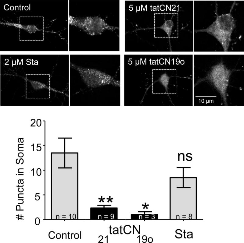
Fig. 5.

CaMKII aggregation within neurons does not require enzymatic kinase activity. CaMKII FingR transfected neurons were imaged in the presence of tatCN21 (5 μM; n=9), tatCN19o (5 μM; n=3), staurosporine (Sta; 2 μM; n=8), or no inhibitor (control; n=10). The top panels show representative images of cells in all four conditions 2 minutes after glutamate/glycine stimulation. Puncta – indicating aggregation – can clearly be seen in the control and staurosporine conditions, but not in either of the tat-peptide conditions. The images were quantified by counting the number of somal puncta, as defined by objects greater than 2 pixels in size whose intensity was greater than 3 standard deviations from the mean somal intensity at 2 minutes. This value was significantly less than control for tatCN19o and tatCN21, as determined by a One-Way ANOVA with post-hoc Tukey’s test (* p<0.05, ** p<0.01, ns = non-significant as compared to control).
