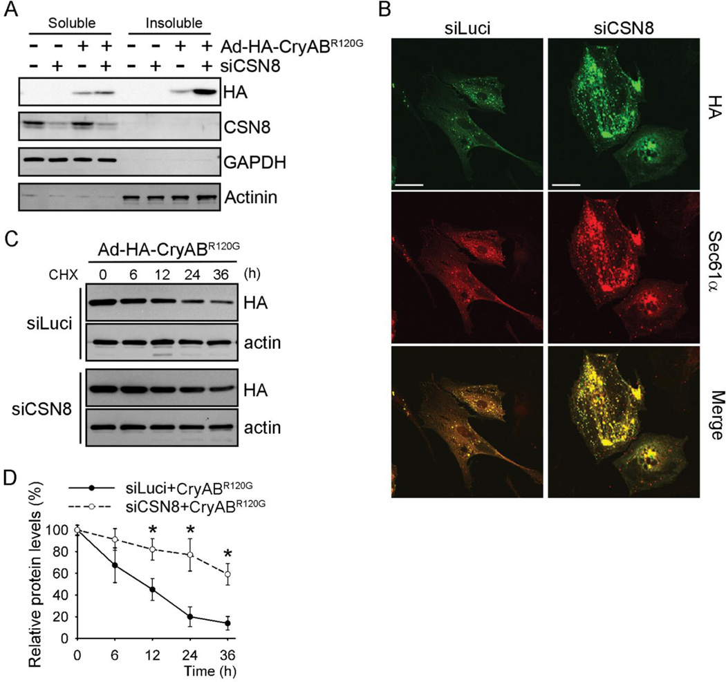
Figure 5. CSN8 knockdown stabilizes CryABR120G in cardiomyocytes.
NRVMs were infected with adenoviruses expressing HA-CryABR120G (Ad-HA- CryABR120G) or β-Gal as indicated. The cells were also transfected with siRNAs against either luciferase (siLuci) or CSN8 (siCSN8). At 72 hours after the siRNA transfection, the cells were harvested for the analyses (A, B) or treated with cycloheximide (CHX, 100 µmol/L) for the indicated times (C). (A) Representative western blot images of indicated proteins in the Triton X-100 soluble and insoluble fraction of cell lysate. GAPDH and α-actinin were probed as loading controls. (B) Immunofluorescent images showing increased protein aggregates in CSN8 knockdown cells. HA-tag (green) and Sec61α(red) were stained for CryABR120G and aggresomes, respectively. Scale bar=50 µm. (C, D) Cycloheximide (CHX) chase assay for HA-CryABR120G. HA-CryABR120G protein levels at the indicated time points were measured using western blot analyses for HA-tag. A representative image (C) and a summary of the relative levels of HA-CryABR120G (D) are shown; *p<0.05 vs. the siLuci+CryABR120G group, n=3 repeats; t-test.

