Figure 7.
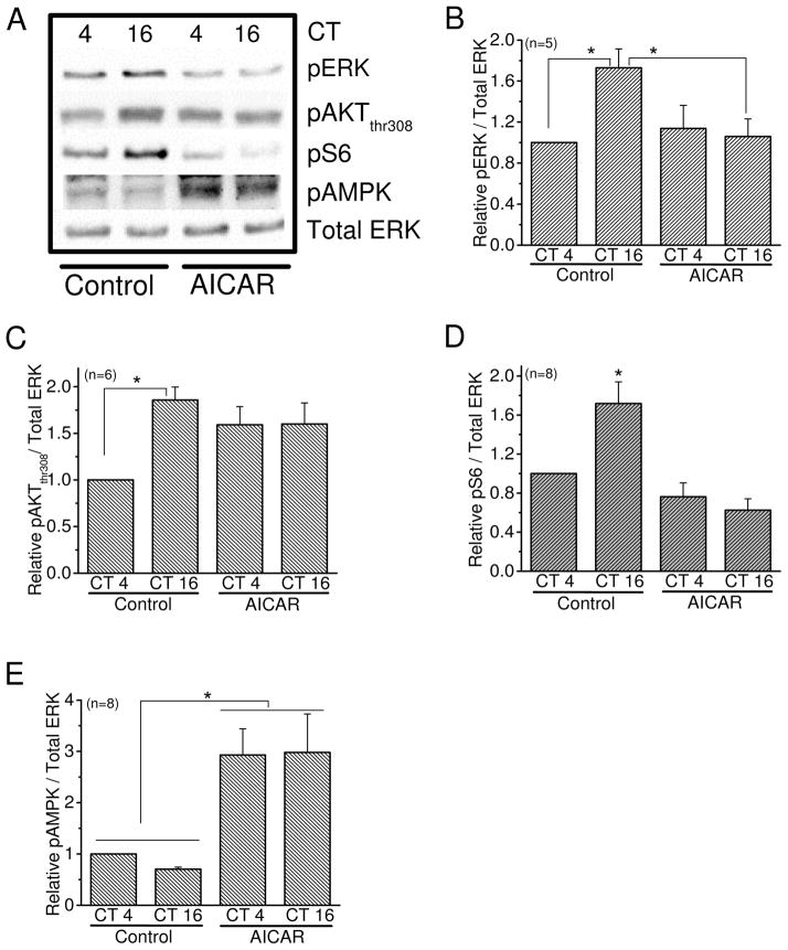
AMPK is a negative regulator of mTORC1 signaling. Chick embryos were LD entrained for 7 days. At E17, the retinas were dissected and cultured in DD. On the second day of DD, the cultured retinas were treated with the AMPK activator AICAR for 2 hr. The retinal samples were collected at CT 4 and CT 16 for immunoblotting analyses. (A) The di-phospho-ERK (pERK), phospho-AKT at Thr308 (pAKT), phospho-S6 (pS6), and phospho-AMPK at Thr172 (pAMPK) were detected from samples treated with AICAR (500μM) or control. Since the total amounts of ERK, AKT, and S6 remained constant throughout the day, total ERK was used as loading control. (B) After treatment with AICAR, pERK is decreased at CT 16 compared to the control group at CT 16. * indicates that the control group harvested at CT 16 is significantly higher than CT 4 of control and the AICAR-treated at CT 16. (C) Treatment with AICAR seemed to increase pAKT at CT 4, but it did not reach statistical significance. * indicates a significant difference between CT 4 and CT 16 of the control groups. (D) The level of pS6 is significantly dampened with AICAR treatments compared to the control group at CT 16. * indicates the control group at CT 16 is significantly higher than the other three groups. (E) The phosphorylation of AMPK served as an internal control showing that treatment with AICAR for 2hr increases AMPK activities. * indicates a significant difference between the AICAR groups and control groups at both CT 4 and CT 16, respectively. * p<0.05.

