
Fig. 6. Abnormal perivascular spaces in Alzheimer's disease.
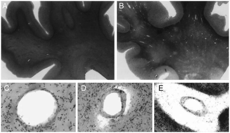
2.5× magnification of hematoxylin and eosin staining of superior frontal gyrus and underlying white matter. A) The white matter of a 74-year old with no CNS diagnosis shows homogenously stained white matter with normal perivascular spaces. B) The white matter of an 80-year old Alzheimer's disease patient contains patches with paler staining and a large number of abnormally enlarged perivascular spaces. 100× magnification of normal C), slightly dilated D) and severely dilated E) perivascular spaces in an Alzheimer's disease patient. Reprinted from [112] with permission.