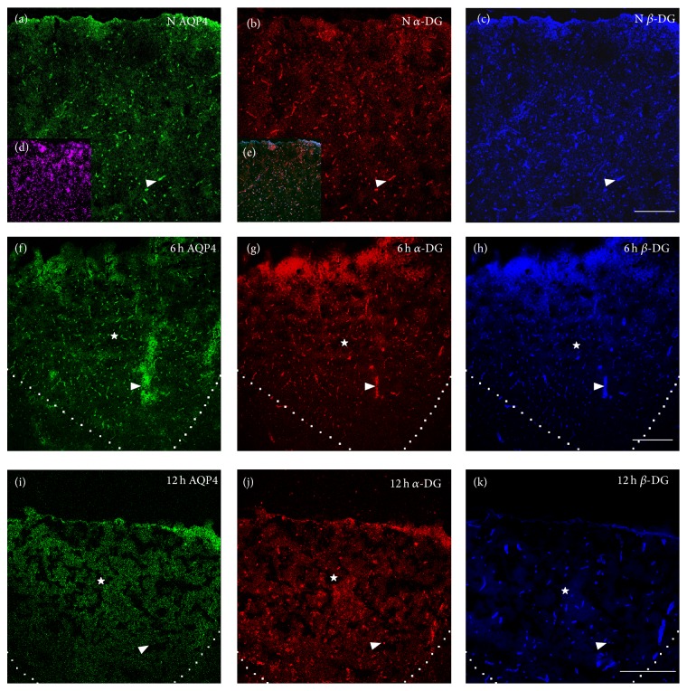
Figure 3.
The expression of AQP4, α-DG, and β-DG in normal brain cortex and in the core lesion at 6 and 12 h after TBI. Green, red, and blue IF signal represented AQP4, α-DG, and β-DG, respectively. Small panels (d) and (e) showed DAPI staining and the merged images, respectively. The area surrounded by dashed line or indicated by star (☆) was the core lesion. Normally, AQP4, α-DG, and β-DG were specially abundant in perivascular endfeet of astrocyte and vessel-like sharp appeared indicated by arrow (△) in (a)–(c). At 6 h after TBI, the vessel-like sharp was maintained and the IF signals of the three proteins were increased in the core lesion shown by (f)–(h). At 12 h, the three proteins were dramatically decreased in perivascular endfeet and punctation-like sharp appeared indicated by arrow (△) and AQP4 and α-DG were lost more seriously than β-DG in (i)–(k). Scale bar: 150 μm.

