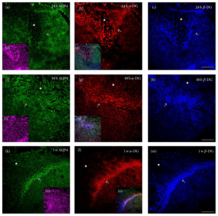
Figure 4.
The changes of AQP4, α-DG, and β-DG expression in the penumbra and core lesion of brain cortex after 24 h of TBI. Green, red, and blue IF signal represented AQP4, α-DG, and β-DG, respectively. Small panels (d), (i), and (n) showed DAPI staining. Small panels (e), (j), and (o) were the merged images. The area surrounded by dashed line or indicated by star (☆) was the core lesion. Arrow (↗) indicated the penumbra of the core lesion. At 24 h, the polarized expression of three proteins in perivascular endfeet was totally lost in the lesion core and the surrounding core area, although α-DG and β-DG were diffusedly increased, markedly, in the lesion core shown by (a)–(c). At 48 h, an obvious penumbra surrounding the lesion core indicated by arrow in (f)–(h) appeared, although AQP4 IF signal was weak but visible. Contrasted with 24 h, at 48 h shown in (f)–(h), the three signals are still lost or diminished in perivascular area. The diffused expression of α-DG was decreased in the lesion core but greatly increased in the penumbra; β-DG continues to be increased in the lesion core and the penumbra; AQP4 was dramatically and diffusedly increased in the lesion core and penumbra (weakly but visible). Until 1 w, the three signals were greatly increased in penumbra but decreased slightly in the lesion core shown in (k)–(m). Scale bar: 150 μm.

